

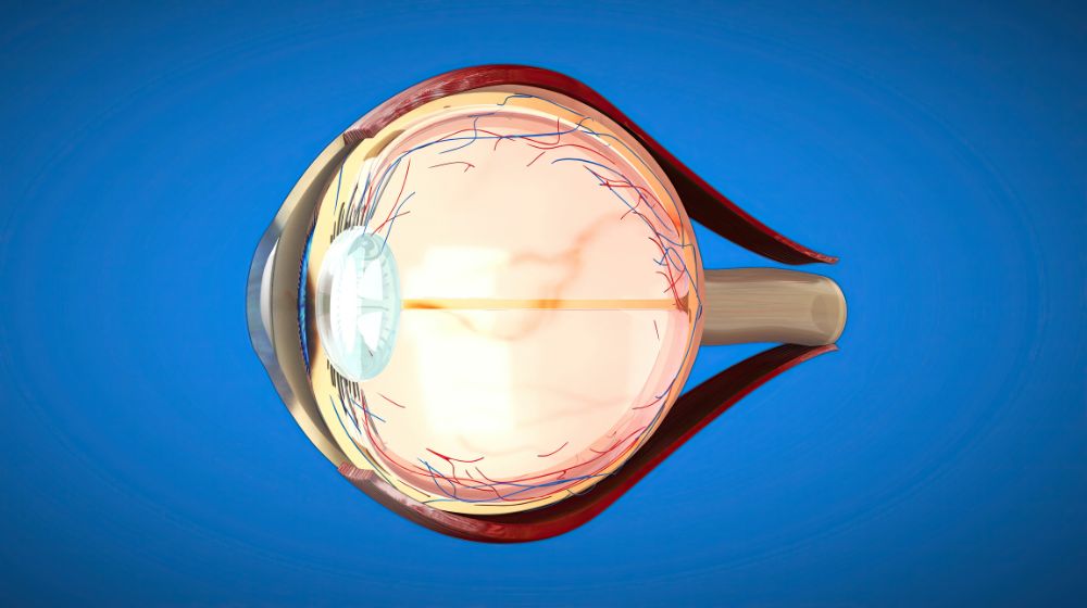
When we talk about vision, everything begins with the layers of retina. This thin but complex tissue at the back of the eye converts light into signals your brain can understand. Without the retina, we couldn’t see shapes, colors, or even light itself.
The retina isn’t just one sheet—it’s made of 10 layers of retina, each with a unique role. From photoreceptors that capture light to nerve fibers that send messages to the brain, every layer is essential. If even one layer is damaged, vision can be affected. That’s why understanding the retina layer of the eye is important not just for medical students, but also for anyone curious about how vision works.
The Structure of Retina Layer of Eye
The retina is located at the back of the eye, lining the inside wall. Despite being less than half a millimeter thick, it contains millions of nerve cells. These cells are organized into layers of retina that work together like a team.
You can think of the retina as a camera sensor. Just as a camera has multiple parts to capture and process an image, the retina’s 10 layers filter, process, and transmit visual information.
The 10 Layers of Retina Explained
Here’s a detailed look at the 10 layers of retina in the order from the outermost to the innermost:
1. Retinal Pigment Epithelium (RPE)
- Acts like a shield protecting photoreceptors.
- Absorbs stray light, preventing blurred vision.
- Stores Vitamin A needed for vision.
2. Layer of Rods and Cones (Photoreceptor Layer)
- Contains photoreceptor cells: rods (for low-light vision) and cones (for color vision).
- This is where light is first captured.
3. External Limiting Membrane (ELM)
- A thin layer that maintains the structure of photoreceptors.
- Acts like a barrier, controlling the exchange of molecules.
4. Outer Nuclear Layer (ONL)
- Holds the cell bodies of rods and cones.
- Essential for transmitting signals from light to nerve pathways.
5. Outer Plexiform Layer (OPL)
- The communication zone where photoreceptors connect with bipolar and horizontal cells.
- Begins the process of contrast detection.
6. Inner Nuclear Layer (INL)
- Contains cell bodies of bipolar, horizontal, and amacrine cells.
- Acts as the “middleman” in visual processing.
7. Inner Plexiform Layer of Retina (IPL)
- Where bipolar cells connect with ganglion cells.
- Important for detecting motion and light changes.
- Plays a big role in sharpening visual signals.
8. Ganglion Cell Layer
- Contains the nuclei of ganglion cells.
- Responsible for carrying signals towards the optic nerve.
9. Nerve Fiber Layer (NFL)
- Made of axons from ganglion cells.
- These fibers bundle together to form the optic nerve.
10. Internal Limiting Membrane (ILM)
- The innermost layer of retina.
- Acts as a boundary between the retina and the vitreous body (the gel inside the eye).
10 Layers of the Retina and Their Functions: Quick Summary Table
| Layer | Function |
| Retinal Pigment Epithelium | Absorbs light, prevents scatter |
| Photoreceptor Layer (Rods/Cones) | Detects light and color |
| External Limiting Membrane | Maintains photoreceptor structure |
| Outer Nuclear Layer | Contains photoreceptor cell bodies |
| Outer Plexiform Layer | Connects photoreceptors with bipolar cells |
| Inner Nuclear Layer | Houses bipolar, horizontal, amacrine cells |
| Inner Plexiform Layer | Connects bipolar cells to ganglion cells |
| Ganglion Cell Layer | Collects signals for optic nerve |
| Nerve Fiber Layer | Forms the optic nerve |
| Internal Limiting Membrane | Boundary between retina and vitreous |
Layers of Retina Mnemonic: Easy Way to Remember
Medical students often struggle to memorize the 10 layers of retina. Here’s a simple layers of retina mnemonic you can use:
“Randy’s Photographs Exhibit Outstanding Outer & Inner Impressive Graphic Nerve Illustrations.”
- R = Retinal pigment epithelium
- P = Photoreceptor layer
- E = External limiting membrane
- O = Outer nuclear layer
- O = Outer plexiform layer
- I = Inner nuclear layer
- I = Inner plexiform layer
- G = Ganglion cell layer
- N = Nerve fiber layer
- I = Internal limiting membrane
This memory trick makes it easier for students preparing for ophthalmology exams.
OCT Layers of Retina: Modern Imaging
In clinical practice, eye specialists use Optical Coherence Tomography (OCT) to see the OCT layers of retina in real-time.
OCT scans produce cross-sectional images of the retina, allowing doctors to detect conditions like:
- Macular degeneration
- Diabetic retinopathy
- Glaucoma
- Retinal detachment
By analyzing the OCT layers of retina, eye doctors can diagnose early changes long before vision loss occurs.
Why the Layers of Retina Are Important
The layers of retina are vital for:
- Vision formation – converting light into signals.
- Color recognition – distinguishing millions of shades.
- Image clarity – filtering out unnecessary signals.
- Eye disease detection – conditions often begin in specific retinal layers.
Even a small defect in one layer, like the inner plexiform layer of retina, can cause significant vision problems. That’s why regular eye checkups at a trusted clinic or eye hospital in Panchkula (for local readers) can help maintain healthy vision.
Common Diseases Affecting Retina Layers
- Macular Degeneration – damages the photoreceptor layer.
- Diabetic Retinopathy – affects blood vessels in multiple layers.
- Glaucoma – damages ganglion cell layer and nerve fiber layer.
- Retinal Detachment – separates retina layers from the back of the eye.
Protecting the Retina Layer of Eye
- Eat foods rich in Vitamin A, C, and Omega-3 fatty acids.
- Protect eyes from UV rays using sunglasses.
- Get regular eye exams with OCT scans.
- Manage systemic conditions like diabetes and hypertension.
FAQs on Layers of Retina
Q1. How many layers are in the retina?
The retina has 10 layers, each with specific functions in vision.
Q2. Which layer of retina contains rods and cones?
The photoreceptor layer (second layer) holds rods and cones.
Q3. What is the inner plexiform layer of retina responsible for?
It processes motion and contrast, connecting bipolar and ganglion cells.
Q4. Can OCT detect problems in retina layers?
Yes, OCT scans clearly show the OCT layers of the retina, helping detect diseases early.
Q5. What is the easiest mnemonic for the layers of the retina?
“Randy’s Photographs Exhibit Outstanding Outer & Inner Impressive Graphic Nerve Illustrations.”
Conclusion
The layers of the retina are a masterpiece of biology. Each of the 10 layers of retina plays a distinct role in helping us see. From capturing light to transmitting signals to the brain, this thin tissue controls the way we perceive the world.
By remembering a simple layer of retina mnemonic, and understanding the 10 layers of the retina and their functions, both students and patients can appreciate just how complex and important the retina is.
Modern imaging techniques like OCT layers of retina further help us protect eye health by detecting problems early. So, whether you’re a medical student, a patient, or just curious, understanding the retina layer of the eye is the first step toward valuing your vision.